Anthropometry comprises a series of noninvasive, inexpensive, and easy-to-performmethods for estimating body composition. However, they are operatordependent and, to be useful clinically, must be performed in a precise,standardized, and reproducible manner. It is recommended that any individualwho performs the measurements should first undergo training to increaseprecision and skill. Without such training, considerable variance willoccur both within and between observers in obtaining and interpretingthe measurements. Standardized methods for collecting anthropometricdata are available and should be utilized.
The anthropometric measurements that are valid for assessing protein-energynutritional status in MD patients include skinfold thickness, midarmmuscle area or circumference, %UBW, and %SBW. An estimate of skeletalframe size is also necessary for evaluating an individual's anthropometricmeasurements. Therefore, a brief description of the methodology andreference tables for evaluating frame size in addition to other measuresare provided.
Skeletal Frame Size
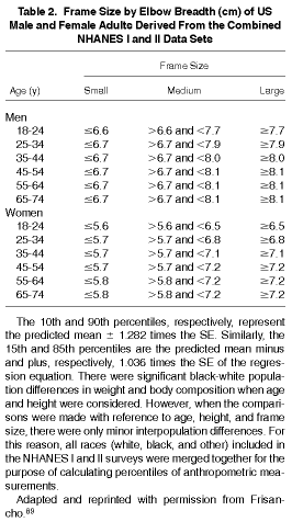
Measurement of elbow breadth is a rough estimate of an individual's skeletal frame size. Frame size estimates of small, medium, and large for males and females are available and presented in Table 2.89
Method for Estimating Skeletal Frame Size
Equipment. Sliding bicondylar caliper.
Method. Ask the patient to stand erect, with feet together facing the examiner. Ask the patient to extend either arm forward until it is perpendicular to the body. Flex the patient's arm so that the elbow forms a 90° angle with the fingers pointing up and the posterior part of the wrist is toward the examiner. Hold the small sliding caliper (bicondylar caliper) at a 45° angle to the plane of the long axis of the upper arm and find the greatest breadth across the epicondylis of the elbow. Measure to the nearest 0.1 cm twice with the calipers at a slight angle (this may be necessary because the medial condyle is more distal than the lateral condyle). An average of the two measurements is used (Table 2).89
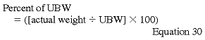
Percent of Usual Body Weight (%UBW)
UBW is obtained by history or from previous measurements. A stableweight in adult dialysis patients may be an indicator of good nutritionalstatus, because adults normally are expected to maintain their bodyweight. The formula below for percent of UBW is appropriate for patientswhose weight has been stable for most of their lives.
Weight loss over time is a simple and useful longitudinal measure tomonitor nutritional status because it is a risk factor for malnutrition.Even if the patient is overweight or obese, a significant weight lossin a short period of time may indicate malnutrition and predict increasedmorbidity and mortality.
Percent of Standard Body Weight (%SBW)
SBW is the patient's actual weight (postdialysis) expressed as a percentageof normal body weight for healthy Americans of similar sex, height,and age range and skeletal frame size.
![]()
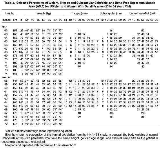
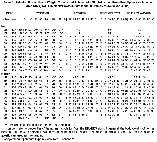
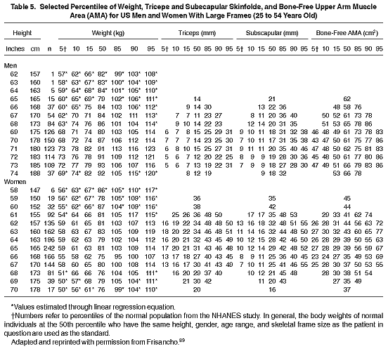
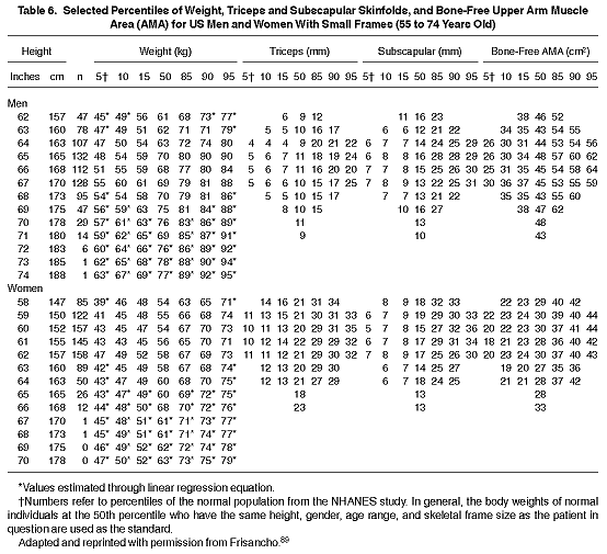
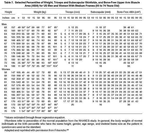
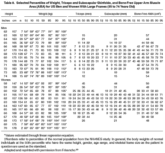
For individuals in the United States, these data are usually obtained from the National Health and Nutrition Evaluation Survey (NHANES). The third and most recent NHANES study indicates that the average American has gained about 7% in body weight.97 This was considered a compelling argument for using the NHANES II data rather than data from NHANES III. However, individuals undergoing MHD who are in the upper 50th percentile or greater of body weight-for-height have an increased odds ratio for survival.97 Patients who are less than 90% of normal body weight are considered to be mildly to moderately malnourished, and those who are less than 70% of normal body weight are considered severely malnourished.85 Individuals who are 115% to 130% of SBW are considered mildly obese, those between 130% and 150% are moderately obese, and those above 150% of SBW are considered to be severely obese.259 Therefore, it is recommended that a target body weight for maintenance dialysis patients is between 90% and 110 % of SBW. At present, it is recommended that the NHANES II data be used for the reference source (Tables 3 through 8).89
Body Mass Index (BMI)
BMI is a useful and practical method for assessing the level of body fatness. BMI is calculated by dividing weight (in kilograms) by height squared (in meters). Based on epidemiological data,85 it is recommended that the BMI of MD patients be maintained in the upper 50th percentile, which would be BMIs for men and women of at least approximately 23.6 and 24.0 kg/m2, respectively. Notwithstanding the greater unadjusted survival data for men and women in the upper 10th percentile of body weight for height,15,85 the large numbers of epidemiological data in normal individuals suggest that persons who are severely obese (eg, %SBW greater than 120 or BMI greater than 30 kg/m2) should be placed on weight reduction diets. Shorter survival also suggests that obese MD patients should also be placed on weight reduction diets, but no studies have been performed in MD patients to determine the safety and efficacy of this theory.
Skinfold Thickness
Skinfold anthropometry is a well-established clinical method for measuring body fat.260 Subcutaneous fat measurement is a rather reliable estimate of total body fat in nutritionally stable individuals. About one-half of the body's fat content is found in the subcutaneous layer.83 Measurement of skinfold thickness at only one site is a relatively poor predictor of the absolute amount of body fat and the rate of change in total body fat because each skinfold site responds differently relative to changes in total body fat.83 Measuring skinfold thickness at four sites (triceps, biceps, subscapular, and iliac crest) that quantify subcutaneous adipose tissue thickness on the limbs and trunk can make an accurate assessment of body fat.86,261,262 Equations have been developed for estimating total body fat from these skinfold thicknesses,260 although these equations have been developed from people without renal failure. Tables 2 through 7 give normal values for triceps and subscapular skinfold thicknesses.89 Nonetheless, measuring skinfold thickness should be considered a semiquantitative measure of the amount or rate of change in total body fat.
In a study that measured four-site skinfold anthropometry, a reduction in percent total body fat was observed in a group of MHD patients when compared with controls.261 Loss of fat from subcutaneous stores occurs proportionally. Therefore, repeated measures in the same patient over time may provide useful information on trends of fat stores.83
Methods for Performing Skinfold Thickness
Measuring Upper Arm Length
Equipment. Flexible, nonstretchable (eg, metal) tape measure.
Method. (1) Ask the patient to stand erect with his/her feet together. (2) Stand behind the patient. (3) Ask the patient to flex his/her right arm 90° at the elbow with the palm facing up. (4) Mark the uppermost edge of the posterior border of the acromion process of the scapula with a cosmetic pencil. (5) Hold the tape measure at this point and extend the tape down the posterior surface of the arm to the tip of the olecranon process (the bony part of the mid-elbow). (6) Keep the tape in position and find the distance halfway between the acromion and the olecranon process that is the midpoint of the upper arm. (7) Mark a (+ ) at the midpoint on the posterior surface (back) of the arm. (8) Mark another (+ ) at the same level on the anterior (front) of the arm.
Measuring Skinfold Thickness (Biceps, Triceps, Subscapular, and Iliac Crest)
Equipment. Skinfold calipers.
Method: triceps skinfold (TSF). (1) Ask the patient to stand with his/her feet together, shoulders relaxed, and arms hanging freely at the sides. (2) Stand to the patient's right side. (3) Locate the point on the posterior surface of the right upper arm in the same area as the marked midpoint for the upper arm circumference. (4) Grasp the fold of skin and subcutaneous adipose tissue gently with your thumb and forefingers, approximately 1.0 cm above the point at which the skin is marked, with the skinfold parallel to the long axis of the upper arm. (5) Place the jaws of the calipers at the level that has been marked on the skin with the marking pencil. The jaws should be perpendicular to the length of the fold. (6) Hold the skinfold gently and measure the skinfold thickness to the nearest 1 mm. (7) Record the measurement. If two measurements are within 4 mm of each other, record the mean. If the measurements are more than 4 mm apart, take four measurements and record the mean of all four.
Method: biceps skinfold. (1) Follow the same procedure as for the TSF, but with the measurement of the biceps skinfold at the front of the upper arm (instead of the back, as with the triceps). The level is the same as for the triceps and arm circumference, and the location is in the midline of the anterior part of the arm. (2) Ask the patient to stand with his/her feet together, shoulders relaxed, and arms hanging freely at the sides. (3) Stand behind the patient's right side. (4) Rotate the right arm so that the palm is facing forward. (5) Locate the point on the anterior surface of the right upper arm in the same area as the marked midpoint for the upper arm circumference. (6) Grasp the fold of skin and subcutaneous adipose tissue on the anterior surface of the upper arm, in the midline of the upper arm, and about 1.0 cm above the marked line on the middle of the arm. (7) Measure the skinfold thickness to the nearest 1 mm while you continue to hold the skinfold with your fingers. (8) Record the measurement. If two measurements are within 4 mm of each other, record the mean. If the measurements are more than 4 mm apart, take four measurements and record the mean of all four.
Method: subscapular skinfold. (1) Ask the patient to stand erect, with relaxed shoulders and arms. (2) Open the back of the examination gown or garment. (3) Palpate for the inferior angle of the right scapula. (4) Grasp a fold of skin and subcutaneous adipose tissue directly below (1.0 cm) and medial to the inferior angle. This skinfold forms a line about 45° below the horizontal, extending diagonally toward the right elbow. (5) Place the jaws of the caliper perpendicular to the length of the fold, about 1.0 cm lateral to the fingers, with the top jaw of the caliper on the mark over the inferior angle of the scapula. (6) Measure the skinfold thickness to the nearest 1 mm while the fingers continue to hold the skinfold. (7) Record the measurement. If two measurements are within 4 mm of each other, record the mean. If the measurements are more than 4 mm apart, take four measurements and record the mean of all four.
Method: suprailiac skinfold. (1) Ask the patient to stand erect, with feet together and arms hanging loosely by the sides. If necessary, arms may be abducted slightly to improve access to the site. This measurement can be taken in the supine position for those unable to stand. The suprailiac skinfold is measured in the midaxillary line immediately superior to the iliac crest. (2) Palpate for the iliac crest. (3) Grasp the skin at an oblique angle, just posterior to the midaxillary line below the natural cleavage lines of the skin. Align the skinfold inferomedially at 45° to the horizontal. (4) Gently apply the caliper jaws about 1 cm from the fingers holding the skinfold. (5) Record the skinfold to the nearest 0.1 cm. If two measurements are within 4 mm of each other, record the mean. If the measurements are more than 4 mm apart, take four measurements and record the mean of all four.
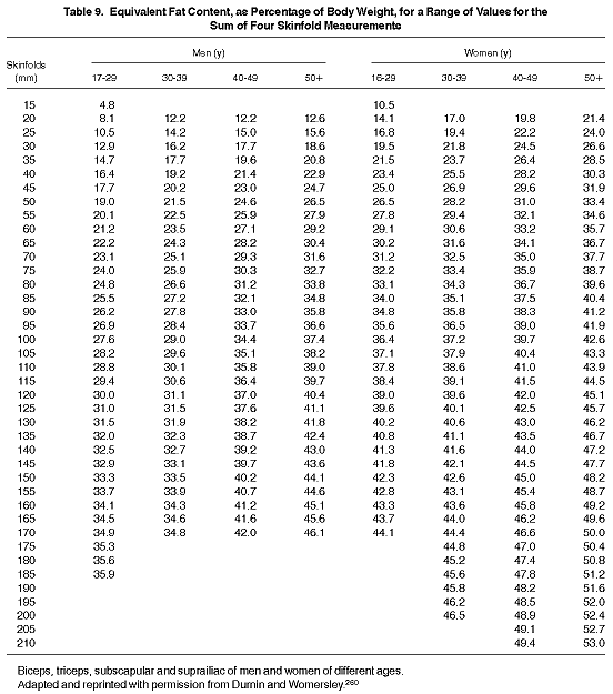
The suprailiac skinfold, as well as the biceps skinfold, may be moreuseful in the research setting than in most clinical settings. It maybe more difficult to obtain the suprailiac skinfold than the other skinfoldmeasurements due to the potential reluctance of patients to expose thatsite. However, the Tables 9 and 10 are provided for those who may wishto incorporate these measurements as a component of the anthropometricassessment of MD or CRF patients. The method for estimating body fatfrom these four skinfold measurements is shown below.
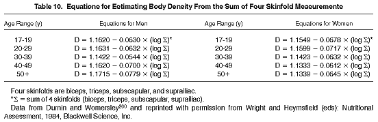
Estimating Body Fat and Fat-Free Mass According to the Method of Durnin and Wormersley260
Method. (1) Determine the patient's age and weight (in kilograms). (2) Measure the following skinfolds (in millimeters): biceps, triceps, subscapular, and suprailiac. (3) Compute the sum (S) by adding the four skinfolds. (4) Compute the logarithm of the sum (S). (5) Apply one of the equations from Table 10 (age- and sex-adjusted) to compute body density (D, g/mL). (6) Fat mass is calculated as follows:
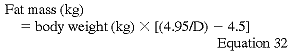
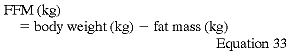
where D is obtained from the formulas shown in Table 10. (7) Fat-freebody mass (FFM) is calculated as follows:
Mid-Arm Muscle Area, Diameter, and Circumference
Anthropometric measures of skeletal muscle mass are an indirect assessment of muscle protein. Approximately 60% of total body protein is located in skeletal muscle--the body's primary source of amino acids in response to poor nutritional intake.83
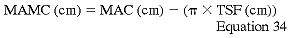
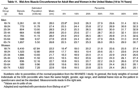
Estimates of muscle mass in an individual, for comparison with a reference population, eg, NHANES, is made by measuring the arm at the midpoint from the acromion to the olecranon. From measurements of both the mid-arm circumference (MAC) and the triceps skinfold (TSF), a calculated estimate of the mid-arm muscle circumference (MAMC) (that includes the bone) can be made using the following formula (Table 11)83:
A more accurate assessment of muscle mass is obtained by estimating bone-free arm muscle area (AMA). Corrected AMA may be calculated from TSF thickness and MAC using the following formulas263:
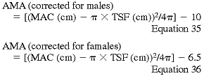
AMA estimates may be inaccurate in obese and elderly subjects (Tables 3 through 8).89
Methods for Performing Mid-Arm Muscle Area, Diameter, and Circumference
Equipment. Flexible, nonstretchable (eg, metal) tape measure.
Method. (1) Ask the patient to stand with his/her elbow relaxed, with the right arm hanging freely to the side. (2) Place the tape around the upper arm, directly over the pencil mark at the midpoint on the posterior aspect (back) of the upper arm. Keep the tape perpendicular to the shaft of the upper arm. (3) Pull the tape just snugly enough around the arm to ensure contact with the medial side of the arm and elsewhere. Make sure that the tape is not too tight that it causes dimpling of the skin. (4) Record the measurement to the nearest millimeter. (5) Check to see if the two measurements are within 0.4 cm of each other. If they are not, take two more measurements and record the mean of all four.