(Adapted from Pediatric Nutrition Handbook [ed 4]. Committee on Nutrition, American Academy of Pediatrics. Elk Grove Village, IL, American Academy of Pediatrics, 1998, pp 168-174.)
Recumbent Length
Measured in children up to approximately 24 months of age or in older children who are unable to stand without assistance.
Equipment. Infant stature board with a fixed headboard and a moveable footboard positioned perpendicular to the table surface, and a rule along one side; pen and paper for recording. Two persons are necessary: one to hold the head and another to measure.
Procedure. (1) The infant may be measured in light clothing, without foot coverings. (2) Place the infant on the table, lying on his back. (3) Hold the crown of the infant's head and bring it gently in contact with the fixed headboard. Align the external auditory meatus and the lower margin of the eye orbit perpendicular to the table. (4) While the head remains in contact with the headboard, a second measurer grasps one or both feet at the ankle. (5) Move the footboard close to the infant's feet as the legs are gently straightened. Bring the footboard to rest firmly against the infant's heels, making sure the toes point straight upward and the knees are pressed down on the table. (6) Read the markings on the side of the measuring board and record the value to the nearest 0.1 cm.
Height
Measures the child who is able to stand unassisted.
Equipment. Fixed measuring device attached to a wall (stadiometer); block squared at right angles or moveable head projection attached at right angle to the board; pen and paper for recording.
Procedure. (1) Have the child remove his or her shoes and stand on the floor, facing away from the wall with heels together, back as straight as possible, arms straight down; heels, buttocks, shoulders, and head touching the wall or vertical surface of the measuring device. A family member or other measurer may be necessary to hold the child's ankles and knees steadily in place. The child's axis of vision should be horizontal, with the child looking ahead and the external auditory meatus and lower margin of the orbit aligned horizontally. (2) Place the head projection at the crown of the head. (3) Hold the block steady and have the child step away from the wall. (4) Note the measurement, and record it to the nearest 0.1 cm. (5) Perform three measurements which are within 0.2 cm of each other and use the average of the three for the final value.
Weight Using an Infant Scale
Equipment. Infant scale that allows infant to lay down; pen and paper for recording.
Procedure. (1) Ask the mother to undress the infant. (2) Place a clean paper liner in the tray of the scale. (3) Calibrate the scale to zero. (4) Lay or sit the infant in the tray. (5) Read the weight according to the type of scale. Make sure the infant is unable to touch the wall or surrounding furniture. (6) Record the weight to the nearest 0.1 kg.
Standing Weight
Equipment. Scale; pen and paper for recording.
Procedure. (1) The child should be weighed in light clothing without footwear. (2) Assist the child onto the platform of the scale. (3) Calibrate the scale to zero. (4) Instruct the child to stand in the center of the platform with feet flat and heels touching, as erect as possible. (5) If using a beam scale, adjust the beam of the scale with the main and fractional poise as necessary until the beam swings freely and comes to rest parallel to the scale platform. Activate the digital scale, if this is the scale used. (6) Read the measurement from the scale, looking squarely at the increments rather than from an angle. (7) Record the weight to the nearest 0.1 kg.
Head Circumference
Measured in children up to 36 months of age.
Equipment. Firm, nonstretchable measuring tape; pen and paper for recording.
Procedure. (1) Have the person assisting hold the infant so that the head is upright. (2) Locate the occipital bone at the back of the head, also the supra-orbital ridges. (3) Apply the tape firmly around the head just above the supra-orbital ridges at the same level on both sides to the occiput. Move the tape up or down slightly to obtain the maximum circumference. The tape should have sufficient tension to press the hair against the skull. (4) Record the measurement to the nearest 0.1 cm.
Mid-Arm Circumference
NOTE: The tables of normal values for MAC and triceps skinfold (TSF) use the right arm. The nondominant arm or arm without hemodialysis access can also be used. Consistent use of the same arm is the most critical factor.
Equipment. Firm, nonstretchable measuring tape; pen and paper for recording.
Procedure. (1) Position at the time of measurement: The mother or substitute sits comfortably on a chair. The child is held, facing forward, by the mother on her lap. The child's right hand is grasped gently but firmly by the mother's hand and placed on the child's hip so that the child's elbow is flexed at about a right angle. Older children who are cooperative need not be held. (2) Briefly explain the purpose of the measurement. (3) Have the mother or substitute bare the child's arm and shoulder. (4) Sit or stand so that the child's arm is relaxed with the elbow point and shoulder facing the measurer. (5) Place the zero end of the measuring tape at the acromium process of the scapula of the arm being measured. Measure to the olecrenon (elbow tip) and note the midpoint. Place a pen mark at the midpoint. (6) Measure around the arm at the level of the mark, with firm and uniform contact with the skin surface. Do not compress the soft tissue of the area. (7) Read the value on the tape and record to the nearest 0.1 cm.
Triceps Skinfold Thickness
Equipment. Lange or other skinfold caliper; firm, nonstretchable measuring tape; pen and paper for recording.
Procedure. (1) Briefly explain the purpose of this measurement. Demonstrate how the caliper is used by applying the jaws to your finger, mother's finger, or the child's finger if possible. (2) This measurement directly follows the MAC measurement. The positioning is the same. (3) Align a long pencil or equivalent directly up the back of the upper arm from the elbow point. Mark along this line at the region of the MAC mark previously made. The two lines should cross at a right angle. (4) The child's arm should be relaxed and hanging at his side. Gently but firmly grasp the fold of skin and subcutaneous adipose tissue approximately 0.1 cm above the point at which the skin is marked, with the skinfold parallel to the long axis of the upper arm. Do not pinch underlying muscle, only the skin. (5) Lift the fatfold enough to clear it from underlying tissue felt deeply with your fingertips. Flex the child's arm to make sure the muscle tissue is not being pinched. (6) Depress the lever of the calipers gently so that the jaws separate. Apply the jaws just below the pinch to the part of the fatfold at the midpoint (defined in number 3) at the same depth as the pinch but about 1 cm down the arm. The jaws should be perpendicular to the length of the fold. (7) Remove the caliper, keeping the left thumb and index finger in position. (8) Repeat the procedure two more times, or until 3 measurements agree within 0.2 mm; record to the nearest 0.1 mm.
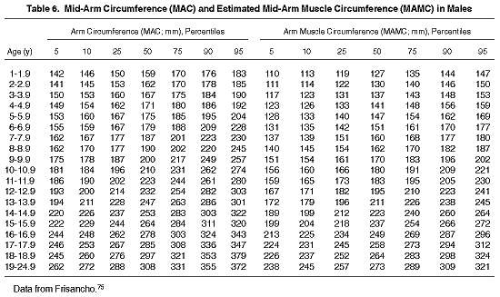
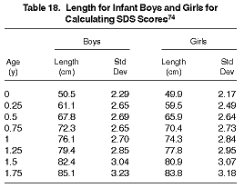
Evaluation of these measurements is done by determining percentiles and comparing them with values from healthy children of the same chronological age and sex, because there are no separate agreed-on standards for growth in children on MD at this time. Standardized growth charts74 and normal tables75 provide the reference data for comparison (Tables 6 through 18). One-time measurements reflect size, whereas serial measurements are necessary for the assessment of growth.

The following are plotted on growth charts on the appropriate graph: standing height or recumbent length, weight, weight for height, and head circumference. Weight for height is determined by plotting the weight and height (or length) measurements on the appropriate grid on the growth chart and noting the percentile. Low weight for height, low height for chronological age, or a low head circumference in proportion to height may reflect chronic nutritional deficits. Parental heights and ethnic backgrounds should be considered when interpreting growth charts.
The mid-arm muscle circumference (MAMC) is calculated from the MAC and TSF measurements according to the following formula:

The mid-arm muscle area (MAMA) can be calculated from the TSF and MAC using the following formula:


The MAC, MAMC, MAMA, and TSF are evaluated according to tables of normal values for children of the same age and sex.75 The most common skinfold thickness measured in children is the TSF because of available normal values and ease of measurement.
Standard Deviation Scores (SDS) are calculated using the patient's actual height compared with control values of the same chronological age and sex (Tables 10 through 17), according to the following equation:

The control subjects used for comparison are of the same chronological age and gender.
SDS for height compares growth rates over specific time intervals.
EXAMPLE: A 7.2-year-old boy has a body weight of 20 kg and a height of 118.5 cm. His SDS for height (Tables 10 and 11) and weight (Tables 14 and 15) are calculated in the following manner:
Height SDS = 118.5 - 122.8 (Table 10)/5.13 (Table 11)
Height SDS = -0.84
Weight SDS = 20 - 23.32 (Table 14)/3.70 (Table 15)
Weight SDS = -0.89
An SDS within two standard deviations encompasses about 95% of healthy North American children; an SDS greater than +2.0 or more negative than -2.0 is associated with either an abnormal increase or decrease in height or weight.15,76
The estimated dry weight can be challenging to ascertain, because weight gain is expected in growing children. Five parameters are helpful in the estimation process: weight, presence of edema, blood pressure, certain laboratory values, and dietary interview. The mid-week, postdialysis weight is used for evaluation purposes in the HD patient, and the weight at a monthly visit (minus dialysis fluid in the peritoneal cavity) is used for the child on peritoneal dialysis. The estimated dry weight is challenging to evaluate in patients prone to edema and must be done in conjunction with a physical examination. Excess fluid may be visible in the periorbital, pedal, and other regions of the body. Edema may affect evaluation of both skinfold and body weight measurements, because expansion in extracellular fluid volume can obscure the effect of altered nutrient intake and metabolism on muscle and adipose tissue mass.76 Hypertension which resolves with dialysis can be indicative of excess fluid weight. Decreased serum sodium and albumin levels may be markers of overhydration. Rapid weight gain in the absence of significant increase in energy intake or decrease in physical activity must be critically evaluated before it is assumed to be dry weight gain.